Deaf to the Thunder, Tuned to the Whisper
For decades, bioelectromagnetics research has been chasing the wrong signal strength.
The assumption was straightforward: stronger magnetic fields should produce stronger biological effects in neural tissue. It turned out to be wrong.
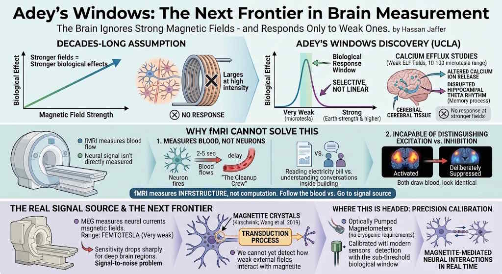
W. Ross Adey and his colleagues at UCLA demonstrated something deeply counterintuitive. Biological effects in the brain display what he called "windows" - specific narrow bands of frequency and amplitude where neural tissue responds. Outside those windows, including at far stronger field intensities, there is no appreciable biological effect at all.
His calcium efflux studies showed that ELF (extremely low frequency) fields at weak amplitudes - in the 10 to 100 microtesla range - altered calcium ion release in cerebral tissue and disrupted the hippocampal theta rhythm associated with memory. The same tissue exposed to stronger fields: no response. The relationship is not linear. It is selective.
This is not a minor detail. It means the brain is not a passive receiver of electromagnetic energy. It is a tuned system - one that filters out the noise of stronger fields and responds only within a specific biological operating range.
Why fMRI Cannot Solve This
Here is what fMRI actually measures: blood.
When neurons fire, blood rushes to the region 2 to 5 seconds later. fMRI detects that blood flow change and calls it neural activity. The neurons have already finished their work. The signal has already traveled. The decision has already been made. And we are standing in the corridor watching the cleanup crew arrive and calling that the event.
Measuring blood flow to understand neural processing is like reading a building's electricity bill to understand the conversations happening inside it. You are capturing the power consumption. Not the content. Not the sequence. Not the mechanism. Just the bill.
It gets worse. fMRI cannot distinguish between excitation and inhibition. A circuit being activated and a circuit being deliberately suppressed look identical - both draw blood. So a regulatory sequence where one region governs and constrains another is completely invisible. You see two lit rooms. You have no idea which one is giving orders.
And for the magnetic field interactions that Adey's Windows point to - the actual electromagnetic computation the brain is running - fMRI is not just inadequate. It is measuring the wrong physical phenomenon entirely.
Blood is infrastructure. It keeps the hardware running. The actual computation is electromagnetic, not metabolic.
Biologists follow the blood. Engineers go to the signal source.
And here is a question no one appears to have asked yet: why build a room-sized magnetic shield to block strong external fields, when you could simply design the sensor itself to only respond within the Adey Window range? Filter out everything above the biological amplitude threshold at the sensor level - mechanically or electronically - and the strong fields never register in the first place. The selectivity is built into the instrument, not the environment surrounding it. This is precisely what the brain already does. It has its own biological filter. The engineering solution is to replicate that filter in hardware.
The Signal Source
MEG - magnetoencephalography - at least measures the right thing: the actual magnetic fields produced by neural currents. These signals are in the femtotesla range, approximately one billion times weaker than Earth's static magnetic field. SQUID sensors in magnetically shielded rooms can detect them. But sensitivity drops sharply for deep brain regions, and the signal-to-noise problem is significant.
What neither fMRI nor current MEG addresses is how the brain's internal magnetic environment - including magnetite crystals identified in human neural tissue by Kirschvink and confirmed by Wang et al. in 2019 - responds to weak external field stimulation within the Adey Window frequencies.
We can measure what the brain produces. We cannot yet detect the transduction process - how weak external fields interact with magnetite to generate neural responses at the biologically active scale.
Where This Is Headed: Precision Calibration
The engineering problem is a calibration problem. SQUID-based MEG sensors exist and function. Optically pumped magnetometers - a newer generation of MEG-adjacent technology - are already operating without cryogenic requirements, making them more flexible and accessible.
The next step is sensors specifically calibrated for the sub-threshold biological window that Adey identified, rather than for Earth-strength or stronger fields. That level of precision would allow researchers to observe magnetite-mediated neural interactions in real time - making visible what has until now only been inferred from indirect metabolic proxies.
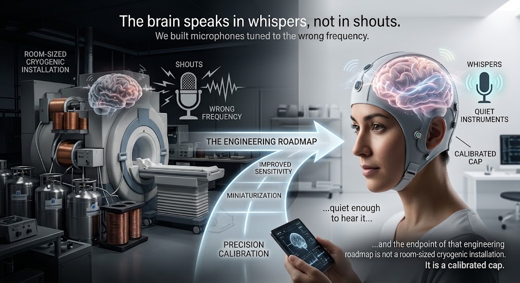
The brain has been telling us for decades that it speaks in whispers, not in shouts. We have spent those decades building microphones tuned to the wrong frequency.
We are finally beginning to build instruments quiet enough to hear it - and the endpoint of that engineering roadmap is not a room-sized cryogenic installation. It is a calibrated cap.